

Extremity lengthening process is achieved by gradual lengthening of mending tissue and soft tissues (skin, muscle, nerves, blood veins etc). This new growth tissue is named as regenerate. Bone and soft tissue are very slowly distracted (pulled apart) from each other so as to be 1 mm per day. If distraction rate is faster than this, bone generation between the edges of the bone that were pulled apart from each other fails and soft tissues such as muscles are left short and tense, nerves can lose functions. If distraction rate is slower than this, early mending can occur between the bone edges and it won’t allow device to lengthen the bone any more.
There are many lengthening devices. The most used ones are the external fixators that hold on to the bone with pin or thicker, screwed nails. There are also devices that are placed fully into the bone. These devices don’t require pin use from outside. Different devices are explained separately.
There are two phases of the lengthening until the time bone fully heals; distraction phase, and consolidation phase. Distraction phase is lengthening phase. When the desired lengthening achieved, newly formed regenerate is weak since there is a low amount of calcium in it. The phase that this bone hardens and calcifies is consolidation.
Lengthening Methods
In our clinic, we use many different devices to distract the bone and soft tissue. Choosing the device is made separately for each case to achieve the desired distraction in the most suitable method for that patient. Generally there are two types of devices; external fixators which hold on to the bone from outside with wires and screwed nails, and internal devices which are placed inside the body, in the place where bone marrow is or on the bone.
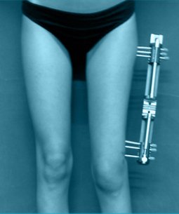
Most known and flexible technique is monolateral (on sided) external fixators (f.e. Orthofix, Heidelberg, EBI) and circular external fixators (f.e. Ilizarov, spatial frames). External fixators can be applied to almost every case. Since external fixator duration becomes too long, we usually combine external fixator with internal nail and prefer (LON: lengthening over nail) method.
We also use fully internally placed, auto-lengthening nails or prosthesis that does not require the use of an external fixator.
Only External Fixator
When only the external fixator is used, fixator needs to stay in place for both the lengthening and consolidation stages. If the device is removed at the end of the distraction phase, newly formed bone would break down and shortening would occur. Therefore external fixator should stay in place until it is determined with roentgens that the regenerate have reached sufficient quality. Frequently, for some more while after the device is removed, the bone is temporarily protected with plaster or orthesis. Total external fixator carrying duration can be calculated for each 1 cm of lengthening is 1 month for children and 1.5-2 months for adults.
Lengthening over Nail
It is a method developed for lowering the duration of external fixator, satisfying the need of protection after removing the device and removing the risk of fracturing the new bone. In this method, as well as the external fixator, metal nail is placed inside the bone. While the nail is placed inside the space bone marrow is, pins of external fixator are put through the bone around the marrow space and nail-pin contact is ensured to not happen. Bone is lengthened as describe in the previous method. After the lengthening is complete, the patient is taken into the surgery room, nail is locked with the holes on it’s both sides and external fixator is removed. In this way, total external fixator duration is lowered from both of distraction and consolidation duration to only distraction duration and made much more shorter. Therefore external fixator carrying duration is lowered by half.
Lengthening Nails and Prosthesis
The most current development are the devices that are placed fully internally and can lengthen the limb without the need of fixator. While these devices remove the infections at the pin location and soft tissue stresses caused by the pin, they also cause less pain and this increases patient comfort. Unfortunately, this method is limited with children and adults who have completed their skeletal development. Lengthening technology is also applied to hip and knee prosthesis. These are most commonly used after bone tumour excisions, congenital deficiencies of femur and tibia.